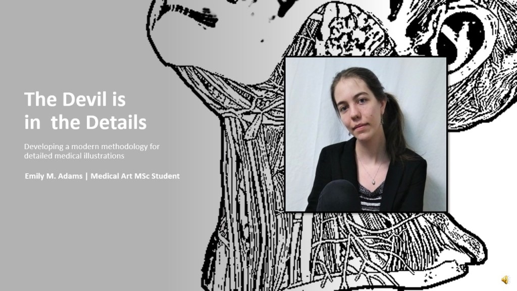
The Devil is in the Details
Developing a modern methodology for detailed medical illustrations
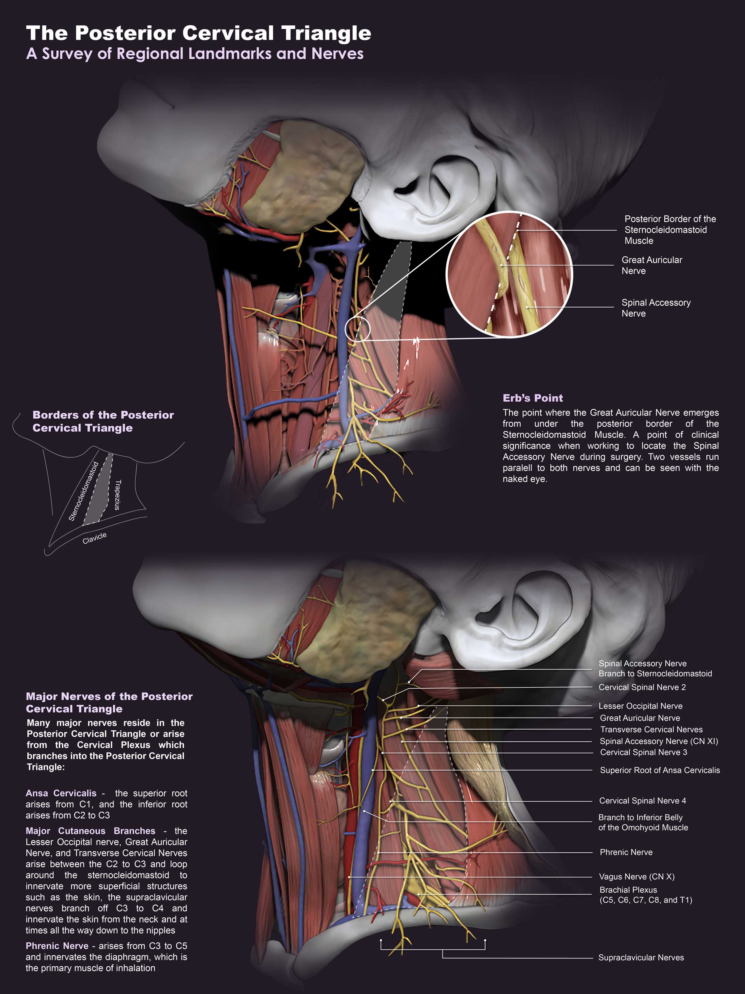
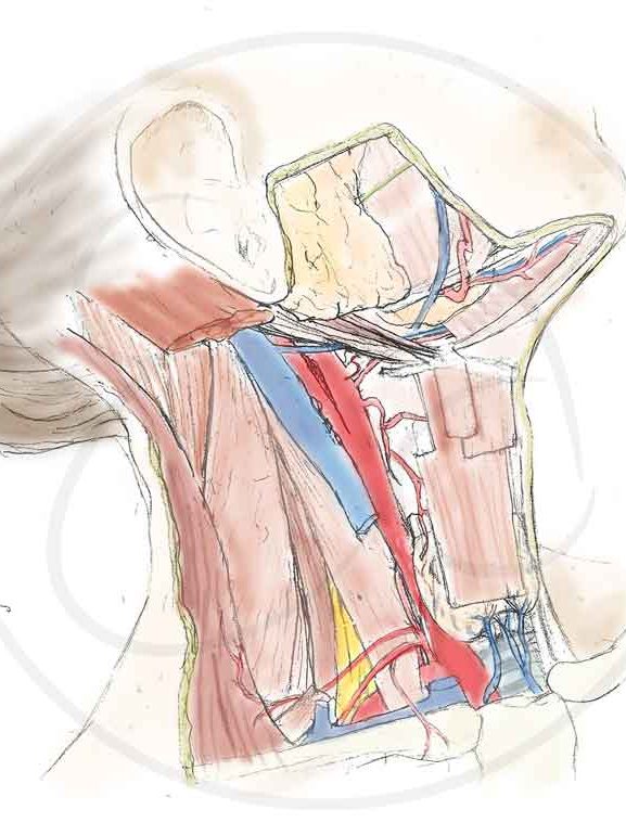
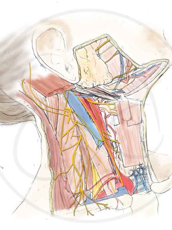
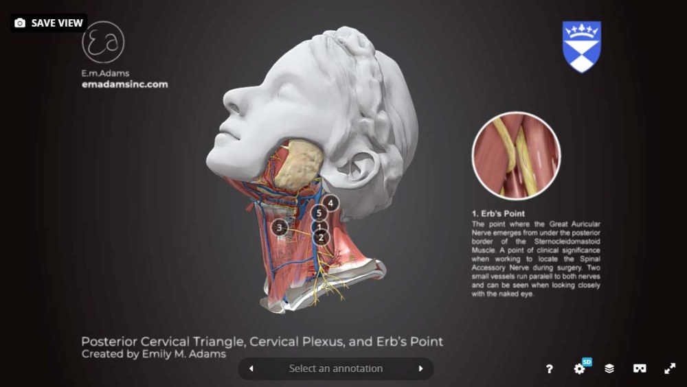
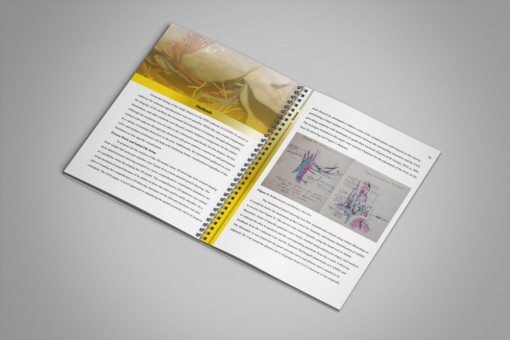
This project aimed to identify successful methods for creating high quality anatomical visualizations through an analysis and comparison of past medical illustrator’s techniques. The creation of 3D models for anatomical education has shown promising acceptance among instructors and students indicating that digital 3D would be a good medium choice for this project. The posterior cervical triangle was chosen as the anatomical region because surgical injury during operations in this area is high, indicating a need for more accurate visualizations of this area. Additionally, the posterior cervical triangle was well-suited to building a 3D model because understanding this anatomical region is dependent on spatial orientation and the spatial relationships between structures. As a part of the survey, participants compared two artistic styles partial grayscale and full color, a 2D and 3D version of the model, and the new visualization to past illustrations. The 3D model can be accessed at https://skfb.ly/6Uwn7. This page shows the process used to create the 3D model and still images produced as a part of this research.
Journal Article
The devil is in the details was published in the Journal of Visual Communication in Medicine in May 2021. The journal article can be accessed at the link below.
Citation
Emily M. Adams & Caroline Erolin (2021) The devil is in the details: developing a modern methodology for detailed medical illustrations, Journal of Visual Communication in Medicine, DOI: 10.1080/17453054.2021.1921566
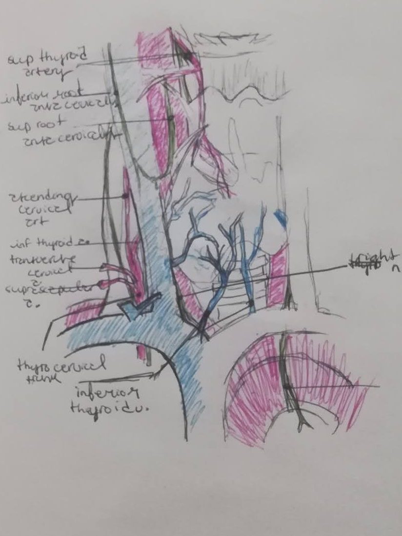
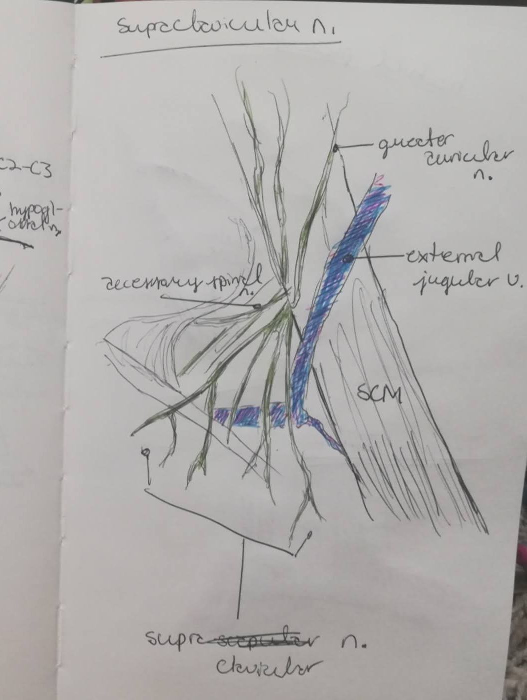
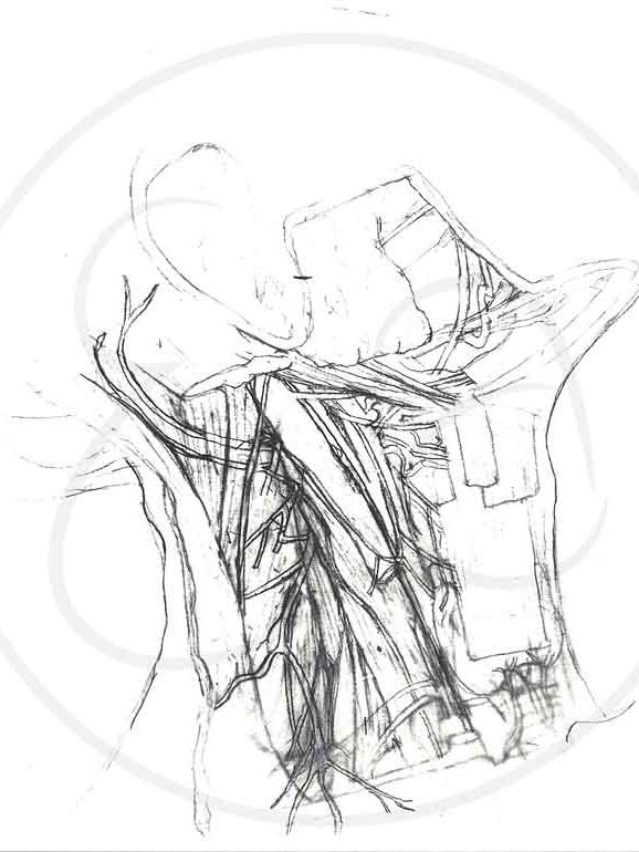
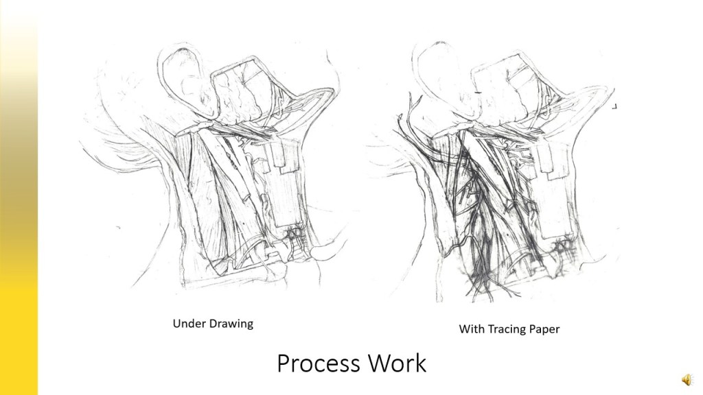
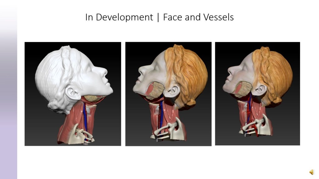
Process Work

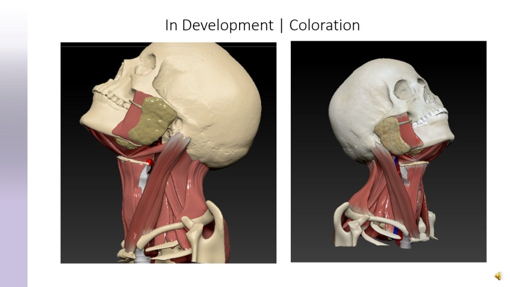
Final 3D Stills


Final Interactive 3D Model
Thesis Layout and Presentation


You must be logged in to post a comment.